List of Chronic Recurrent Multifocal Osteomyelitis (CRMO) Research and Information

This is a collection of research and information on chronic recurrent multifocal osteomyelitis (CRMO). It is not all inclusive. You can search for more studies at PubMed.gov.

Autoinflammatory-Search.org -Chronic Nonbacterial Osteomyelitis (CNO): Chronic Recurrent Multifocal Osteomyelitis (CRMO); and Synovitis, Acne, Pustulosis, Hyperostosis, Osteitis Syndrome (SAPHO) Symptoms and lab results for CRMO and SAPHO on the autoinflammatory database website.

Textbook of Pediatric Rhematology Autoinflammatory Bone Disorders Chapter – This was written by experts Dr. Polly Ferguson and Dr. Ronald Laxer. It has details on clinical symptoms, differencial diagnosis, and treatment for CRMO, Majeed syndrome, DIRA, and cherubism.

Chronic Recurrent Multifocal Osteomyelitis (CRMO) – This is one of the easiest to understand descriptions of CRMO. “Chronic recurrent multifocal osteomyelitis (CRMO) is an autoinflammatory (NOT autoimmune) disorder that mostly affects children. It comprises periodic bone pain, fever, and the appearance of multiple bone lesions that can occur in any skeletal site. The origin of this disease is unclear, but genetics appears to play a role. The clinical and radiological features on the disease are variable and the diagnosis can be difficult.”

Common symptoms include deep aching pain, limping, and fever. Other symptoms can include uveitis, IBD, psoriasis, acne, and pustules on the soles of the feet and palms of the hand. In some cases, Majeed syndrome is associated with CRMO. Other names for CRMO include SAPHO, chronic multifocal osteomyelitis, and chronic recurrent multifocal osteomyelitis.

CRMO Phenotypes Identified in Large French Cohort – Gives detailed information on 3 subgroups of CRMO patients, their disease progression and remission rates.

“In our cohort, after a mean disease duration of 4 years, only 74/171 (43%) of cases of CRMO were considered to be in clinical remission of which 40% were under treatment [while] among CRMO patients with active disease at the last medical visit, 71% remained on therapy [although] some of them had discontinued treatment,”

“These results suggest that CRMO may have a poorer prognosis than previously described with more frequently active disease at follow-up.”

Chronic recurrent multifocal osteomyelitis (CRMO) in children – single-centre clinical observations and experience.-  Study of 42 children diagnosed with CRMO. Median age of onset was 10 to 11 years old. Most had bone pain as the first symptom. During flares, osteocalcin levels were high.

Unusual onset of a case of chronic recurrent multifocal osteomyelitis – “We report here a case a 12 years old girl with CRMO arising with recurrent episodes of left supraorbital headache, followed by the appearance of a periorbital dyschromia. Magnetic resonance imaging (MRI) of the skull and orbits revealed an important subacute inflammatory process. Few months after, the child presented a painful swelling of the left clavicle; the histological examination of the related biopsy allowed to establish the diagnosis of CRMO.”

Autoinflammatory bone disorders with special focus on chronic recurrent multifocal osteomyelitis (CRMO) This is a review of autoinflammatory bone disorders including pyogenic arthritis, pyoderma gangrenosum and acne (PAPA), deficiency of IL-1 receptor antagonist (DIRA), Majeed syndrome, and CRMO. Symptoms, diagnosing, and treatment options are covered. From the article: “Autoinflammatory bone disorders are the result of a disturbed regulation of the innate immune system, resulting in immune cell infiltration of the bone and subsequent osteoclast differentiation and activation, osteolysis and bone remodeling. Though bone biopsies usually remain sterile, lesions mimic infectious osteomyelitis in histology and on radiographs [2-5]. Interestingly, autoinflammatory bone disorders are associated with inflammation of the skin (palmoplantar pustulosis, acne, psoriasis, Sweet syndrome) and/or the intestine (Crohn’s disease, ulcerative colitis, coeliac disease).”

“Furthermore, CNO can be part of the so-called SAPHO syndrome (Synovitis, acne, pustulosis, hyperostosis, and osteitis), which manifests in adolescence or adulthood. Whether CRMO in juvenile patients and SAPHO syndrome are the same disorder in different age groups remains to be determined.”

Chronic Recurrent Multifocal Osteomyelitis in Children: A Multidisciplinary Approach is needed to establish a Diagnosis. – This is a single case report of a 6-year-old girl diagnosed with CRMO. It goes into detail of her symptoms that included fever and swelling of her leg, blood test results, scans, and more. “This study clarifies the clinical and radiologic features of CRMO.”

Chronic recurrent multifocal osteomyelitis – Basic explanation of CRMO from ORPHA.net. Average age of onset is 10 years old. Bone inflammation can occur anywhere, but this notes that: “Metaphyses and epiphyses of the long bones are most frequently affected, in addition to the pelvis, the shoulder girdle and the spine.” Also noted, “Adult-onset forms are reported that resemble SAPHO syndrome. It is not clear whether SAPHO syndrome and CNO/CRMO are two separate entities or if they are part of the same disease spectrum with CNO/CRMO being the pediatric equivalent of SAPHO syndrome.” Most cases have periods of frequent flares and then remission and eventually resolve over time. In some, CRMO causes permanent bone deformities.

Whole-body magnetic resonance imaging: an essential tool for diagnosis and work up of non-oncological systemic diseases in children – “Twenty-one children underwent general anesthesia. WBMRI was a useful tool to provide correct diagnosis in chronic recurrent multifocal osteomyelitis (CRMO), and to identify the origin of fever or arthralgia of unknown etiology.”

Chronic recurrent multifocal osteomyelitis: case series of four patients treated with bisphosphonatesCase series of four patients with CRMO and treatment outcomes. No patients responded to NSAIDs. All were treated with either alendronate or pamidronate with good response. This induced clinical remission in 2 of the 4 patients.

Chronic recurrent multifocal osteomyelitis: typical patterns of bone involvement in whole-body bone scintigraphy. – This study involved performing whole-body magnetic resonance imaging on 53 patients with CRMO. The results show specific common characteristics of CRMO and shows that WB-MRI is an important tool in diagnosing CRMO. “Results: WB-MRI revealed multifocal lesions in all but one patients. Only 26 of them had presented with multifocal complaints. We detected 1 – 27 geographic lesions/patient (mean 9.7). 510 of 513 lesions were significantly hyperintense compared to normal bone marrow. The pelvis, lower extremities, shoulders and spine were most frequently involved. 40 patients (75 %) had bilateral symmetrical involvement of bones. Most of the lesions were located in tubular bones, in 87 % adjacent to one or both sides of a growth plate. 32 % of lesions showed periosteal involvement. Of 456 affected bones, 33 (7.2 %) were deformed, 6 (18 %) were vertebra plana. Conclusion: In the absence of more specific diagnostic criteria, WB-MRI can, in synopsis with clinical findings, substantially contribute to a rapid diagnosis of CRMO.”

Chronic recurrent multifocal osteomyelitis in a 13 year old female athlete: a case report. – In this case, a 13-year-old girl had ankle pain and a history of sore feet, various pains in knees, back, and shoulder along with headaches and sensitive skin. She did not have unexplained fevers. Her father also had a history of unexplained muscle and bone pain. This study includes many other details such as differential diagnosis, treatment options, and MRI pictures.

Diagnostic criteria from this study:

Iyer et al.12, have formulated systematic approach to assist in making the diagnosis of CRMO by exclusion using the following criteria:

  1. Lack of causative organism
  2. No abscess, fistula or sequestra formation
  3. Radiographic appearance of sub-acute or chronic osteomyelitis
  4. Atypical location compared to infectious osteomyelitis
  5. Non-specific histopathologic and laboratory findings compatible with sub-acute or chronic osteomyelitis or other known disease process
  6. Prolonged (> 6 months) and recurrent painful symptoms
  7. Accompanying pustulosis palmoplantaris or acne

The laboratory findings suggest an inflammatory process with evidence of elevated ESR, C-reactive protein and alkaline phosphatase in approximately two-thirds of cases. A tissue biopsy is commonly required in order to rule out more sinister diagnoses such as tumour or infection. Cultures of blood and bone, along with microbial laboratory assays are negative for infectious processes.

Also noted, “CRMO is widely believed to be a pediatric equivalent of SAPHO syndrome.”

Chronic recurrent multifocal osteomyelitis causing an acute scoliosis. A case study of a 15-year-old girl who developed scoliosis secondary to CRMO.

Autoinflammatory bone disorders: update on immunologic abnormalities and clues about possible triggers.

Typical patterns of bone involvement in whole-body MRI of patients with chronic recurrent multifocal osteomyelitis (CRMO). – “In the absence of more specific diagnostic criteria, WB-MRI can, in synopsis with clinical findings, substantially contribute to a rapid diagnosis of CRMO.”

Chronic recurrent multifocal osteomyelitis.Chronic recurrent multifocal osteomyelitis (CRMO) is an autoinflammatory bone disease occurring primarily in children and adolescents. Episodes of systemic inflammation occur due to immune dysregulation without autoantibodies, pathogens or antigen-specific T cells.”

Chronic recurrent multifocal osteomyelitis: experience from a single pediatric rheumatology center over the past ten years

Chronic non-bacterial osteomyelitis in childhood – a comprehensive review.The clinical picture of childhood chronic recurrent multifocal osteomyelitis (CRMO) is characterized by an aseptic chronic osteomyelitis, most often affecting the metaphyses of the long bones. Skin inflammation (palmoplantar pustulosis, psoriatic lesions, acne) and inflammatory bowel disease may be associated with CRMO and therefore subsume this disease into the entity of SAPHO syndrome.”

Chronic recurrent multifocal osteomyelitis mimicking osteoid osteoma. A detailed case report of a 9-year-old girl diagnosed with CRMO. Includes testing done and radiograph images.

Schnitzler’s Disease as an Important Differential Diagnosis of Chronic Recurrent Multifocal Osteomyelitis: A Case ReportSchnitzler’s disease and CRMO can have similar symptoms. “CRMO and Schnitzler’s syndrome are diagnosed by exclusion. At first sight, both diseases can be similar in their manifestation. However, considering the patient as a whole, the age of onset, the male gender, and the very atypical pruritic urticarial lesions on the trunk led to the correct diagnosis. CRMO is associated with pustulosis palmoplantaris, whereas Schnitzler’s disease is associated with neutrophilic dermatitis.”

A case of acute lymphotic leukemia which was difficult to make a diagnosis of CRMO for moving arthralgia and periarticular swelling.

Current Understanding of the Pathogenesis and Management of Chronic Recurrent Multifocal Osteomyelitis – Detailed overview of CRMO including diagnosing, symptoms, treatment, and genetics. Majeed, DIRA, and SAPHO are discussed.

Spinal involvement in chronic recurrent multifocal osteomyelitis (CRMO) in childhood and effect of pamidronate. This study looked at how many with CRMO had involvement of the spine. Of 102 children and teens, 27 had spine deformities, back pain, and/or spinal lesions. Pamidronte was determined to be effective with few side effects.

Chronic Recurrent Multifocal Osteomyelitis-The Prevalence of Lower-Limb and Foot Involvement -“This study presents one of the largest published cohorts of pediatric patients with CRMO and also presents racial/ethnic group data that have not previously been reported in other studies. Despite being a condition considered to affect the metaphysis of long bones, the ankle area and foot bones were also frequently affected.”

Parotid swelling and chronic recurrent multifocal osteomyelitis of mandible in children – “Parotid mass or swelling is a common presentation of CRMO involving the mandible.”

A proposed treatment scheme for chronic recurrent multifocal osteomyelitis (CRMO): a case series of nine patients – “Our treatment policy in CRMO is to start with NSAIDs and MTX. If the patient is refractory to these drugs, anti-TNF treatment is commenced. Anti-TNF treatment was effective in the treatment of our CRMO patients, We suggest that extending the dosing interval is an effective option once they are in remission for 6 months. We also suggest that imaging may be included as an outcome measure in patients with CRMO.”

Like Our Autoinflammatory Chart? You’ll Love What’s Coming Soon!


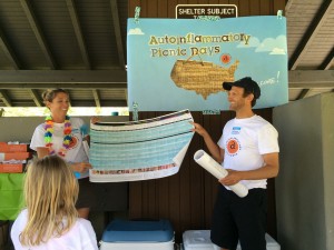
Julie Cunningham, board member & advocate, and Dr Hal Hoffman MD, medical advisor showing off the chart at our 2014 Autoinflammatory Picnic in Southern CA. Photo credit Maria Quintos

Julie Cunningham, board member & advocate, and Dr Hal Hoffman MD, medical advisor showing off the chart at our 2014 Autoinflammatory Picnic in Southern CA. Photo credit Maria Quintos

UPDATE: The Autoinflammatory Search online database is now live! Click here to see it.

Our Comparative Chart of Autoinflammatory Diseases has been available in print and online for a year, and has become a leading reference and educational tool for these diseases in the world.  We had no idea how popular it would be, but this is good news for awareness! The chart has already helped a number of patients get diagnosed, and onto beneficial treatment.  You may have even seen it featured in this recent video about a newly classified autoinflammatory disease, NLRC4-MAS on The National, a major Canadian news show.

Medical professionals like that this expanded comprehensive wall chart has easy to compare information and resources for each disease on it, as prior to the release of the chart, there was no other place to do an easy comparison of all these diseases.

Doctors are using this large chart to not only learn about these diseases, but many have told us that they have studied it for their rheumatology board exams, and have used it to teach other doctors about autoinflammatory diseases. We have handed out thousands of copies at rheumatology conferences worldwide, and have shipped hundreds out by mail to doctors and other medical professionals, in addition to having the chart downloaded online  thousands of times.  If you are wondering how to use it yourself, please read this blog post.

What’s next? The Autoinflammatory Disease Web Application!

With our first debut of the comparative chart at the Autoinflammation 2013 Congress during the abstract and poster presentation session, we were encouraged by leading researchers and experts on autoinlflammatory diseases to take the information and comparative nature of the chart to the next level in a multilingual, digital, interactive and online system.

This is a project that is meant to bring many interested parties from all aspects of these diseases together. Many experts have been asking for us to work on this, and have volunteered to help over the past year, and we are looking forward to working with them on this project to meet the needs of medical professionals, researchers, caregivers and patients.

Why is this needed?

It is not going to be “just another website” but a tool that will transcend our current chart, yet it will still have the ease of use and comparative nature built into a system that will be more functional for digital and mobile uses. The complexity of the web application creation and programming of the search capacities, plus the amount of data to input into this in multiple languages is a monumental task.  But it is possible, and we have assembled a team that will bring this project to pass over the next few months.

This web application will be free and accessible to all online. We are going to collaborate with medical societies, and research centers worldwide on this project to have it linked to not only our organization website, but to all interested and relevant medical societies, clinical centers and other resources.

We have found that most of the current information on these diseases resides in medical journals that are not freely accessible to the general public.  Compiling new information and insights on these diseases, with cited references and links in many languages will help to bridge this informational divide.

Having the web application in a number of languages will help to overcome the lack of information about these diseases in certain parts of the world, which will have a great impact on improving understanding, diagnosis and treatment.  We also hope to develop more patient materials, and also guidelines and resources for medical professionals to use to promote best practices and standards of care and treatment for autoinflammatory diseases.

In addition, we will also have links to all the latest research, clinical trials, genetic testing labs and other helpful information for each disease, and helpful materials for patients all in one site online.  We are working with experts on autoinflammatory diseases, and innovative designers and digital specialists to make this into a very easy to use, helpful tool that can be linked and collaborated on with various doctors, medical centers and medical societies worldwide to make this the “hub for all things autoinflammatory.”

Customized search options will be a key component of the web application

Some ideas that we are working on is how to best show groupings of these diseases, their connections and contrasts. We are also working on a set of   search options to facilitate easy head-to-head comparison between the diseases by symptoms, labs, other clinical findings.

Doctors are clamoring for information on these diseases, in an up to date, easy to use format that will be a “one stop shop” to find all the needed resources, and use as a tool to learn, educate, and help to diagnose patients and fellow doctors about these diseases.

There will be a search option of the main mechanism(s)that cause or mediate the diseases. Or you can compare diseases by the affected protein, gene or chromosome region, so doctors that may be doing new research or even whole exome sequencing that find mutations in certain regions in the DNA can use this information to find out more about diseases that may be associated with certain mutations.

In addition, we will have the images that we have on our chart, but we can have additional photos to show more findings for each disease.  We will also feature links to various medications, and research that discusses experimental care and treatment for these diseases, or direct links for government-approved medications for some of these diseases.

Our present chart will also be refined and expanded and will still be in print as well as a pdf about these diseases. But we have at least four more diseases to add to it, so it is getting to be enormous in print, and as a pdf!  We will likely have to make the chart into two rows of comparative content.

Funding and support

Picture 9

We are pleased to announce that we recently received an unconditional grant from Swedish Orphan Biovitrum AB (Sobi) of $40,000 that is being used to support the development of this web application, and we cannot thank them enough!  Sobi is a leader in rare disease treatments, and has seen the need and potential for this web application that will help spread more awareness for autoinflammatory diseases, and aid in the diagnosis of patients.

More awareness and focus on autoinflammatory diseases will also lead to more interest for research for these conditions as well.  This generous grant from Sobi will help this project to become a reality much sooner than anticipated, which is amazing! Truly, we are so thankful and deeply touched by their support for this project, and also to our donors from the patient community, the general public and industry that are helping us on many levels.

Currently, we are seeking additional funding from a number of other sources to also help with the project, and to help this effort to grow and be sustained for many years. We are filing for a few grants and will announce other support as soon as it is available.

Please contact us or email directly to karen@autoinflammatory.org  if you have an interest in helping to give us insights on what you would like to see with this web app, if you want to support the project in any way from translation, content development, collaborative efforts or grant funding.

The initial costs to develop this web application are higher than many other projects that we have done, but it will have the largest impact for years to come, and will not require lots of funding to sustain over time.  These funds will be focused on this process, and we are also relying on volunteer medical professionals and experts to help with this project as well, which will help to preserve resources for this project. The majority of the cost for this project is going towards programming, development, and data management.

What matters most

The purpose of all our awareness efforts is our strong belief that free access to helpful information and educational materials can help to aid in earlier, and more accurate diagnosis that can lead to better care and treatment for patients that can greatly improve their life and health.

Our Inspiration

Our patients, their families and the doctors and researchers caring for autoinflammatory diseases are our greatest motivation for this project, and they are asking for us to take this to another level.

We have heard from a number of patients in the past year that have directly benefited from having our materials and resources available to their doctors, or themselves online. They email or share their experiences in our private online patient communities, and many have started helpful treatments that have greatly improved their health, and quality of life.This is what motivates us, and why this project matters.

There are likely many undiagnosed patients suffering in the world with these diseases right now, and diagnosis is the first step to getting them help.  In addition, we do help to refer patients to clinical trials, and centers that can help them as needed, and we have had a surge of requests over the past year, thanks in part to our comparative chart and other materials in print, and online.

There is nothing else out there like this! (Which is intimidating, but ripe with potential)

Currently, there is no other unified source to find all the information about systemic autoinflammatory diseases, with a database of symptoms that can be searched and compared head to head or my symptoms with links to numerous references, genetic testing laboratories, clinical trials, and treatment options, in addition to content for patients to use that is in many languages.  There are a number of resources online, including our website (that features the chart and detailed content for a number of these diseases) but the number of diseases that are now autoinflammatory has expanded greatly, so we will be updating our website as well over the next year as well.

Surprisingly, there are very few interactive scientific charts and sites that we have found so far that come close to what is being developed with this project. But, after the many hours that went into making our first comparative chart I realized quickly why nobody had ever made such a chart of all the known systemic autoinflammatory diseases before us! It is a lot of work, and a labor of love!

At the time I was developing the chart content and references for the experts to edit, I was doing a lot of the work on my laptop, at the bedside of my child that was in the hospital with complications with his autoinflammatory disease, in between my part time work as an RN at another hospital and running our org. That month was a bit of a blur, since we were also in the middle of a move, but we made it all work. He is my inspiration, in addition to all the other patients, and our hope is that more people can be helped from our efforts. I never imagined that chart would have this big of an impact, but we are very pleased that it has so far led to more diagnosis, and understanding of these diseases. This next phase will be even more work, but has even more potential to help even more people. Someday, I will get back to doing photo albums, crafts, sewing cross stitch and other projects in my “spare time” but this is what needs to happen now!

Our comparative chart has been nicknamed the “periodic table of autoinflammatory diseases” so I started to look for a digital version of the periodic table of the elements. This dynamic periodic table of the elements has an interactive, multi-layered digital chart format that was inspiring to me, and I truly appreciate the effort that was put into it! But we quickly realized that we needed to take this in a different direction, and away from a literal chart format.

We started to consult digital designers and programmers on this project, to get an estimate of costs and what was possible, and found that there is really very little so far using technology in the way that we have envisioned. So, this will not only be an essential resource for doctors and patients, but may also be an innovation for how to deal with comparing diseases and curating resources.  This potential to not only solve a need for autoinflammatory diseases, but to also help innovate medical information and how it is delivered to people has captured the interest of a number of experts.

Some good examples of existing searchable medical sites that have some content for a few of these diseases are immunology: immunodeficiency search and findzebra.  These are very good, but unfortunately, you cannot click on words in the main copy to “go deeper” into the content, or go to helpful medical journals clinical trials, patient resources or a number of other things.  In addition, we found that only a few autoinflammatory diseases are represented on these search tools, and on findzebra, the sources of content that the search engine found on the internet for some of these diseases had not been updated in over 10 years.  So much has been discovered in that time period that there is a great need for more up to date, focused information and resources.

Hopefully our web application will launch in mid 2015, and we hope that our efforts and the capacity of modern technology can help us to achieve what we are trying to do with this new project.  It will be a work in progress for years to come, and we are so thankful for the support of our donors, grantors, and medical experts that believe in this project. We would not be able to even consider this without them.  Thank you!

List of Systemic-Onset Juvenile Idiopathic Arthritis Research and Information

Updated 2/16/18

Systemic-onset juvenile idiopathic arthritis is one type of juvenile arthritis. SoJIA/SJIA has some differences from other subtypes due to it being autoinflammatory and systemic in nature. Below is a list of studies and other information on diagnosing and treating SoJIA.

Systemic JIA Foundation – Lots of great information on SJIA and resources for SJIA families at this website.

SJIA Parents’ Questions Answered by Doctors at Family Day – Parents ask questions at the SJIA Family Day in Cincinnati in 2017. Read the answers from the expert doctors here.

Systemic Arthritis in Children: A Review of Clinical Presentation and Treatment An comprehensive overview of SoJIA. This paper covers age of onset, symptoms, complications, prognosis, and treatments.

Genetic architecture distinguishes systemic juvenile idiopathic arthritis from other forms of juvenile idiopathic arthritis: clinical and therapeutic implications

“The lack of shared genetic risk factors between sJIA and other JIA subtypes supports the hypothesis that sJIA is a unique disease process and argues for a different classification framework. Research to improve sJIA therapy should target its unique genetics and specific pathophysiological pathways.

At a time when an emphasis is being placed on the personalization of medicine, it is important that we move away from broad classifications based on non-specific clinical observations and move towards the use of molecular and genetic data in establishing diagnoses, as well as pathophysiology. In turn, clinical practice will advance as these data are translated into targeted therapeutic approaches.

Perhaps it is time to separate this condition from JIA all together to make clear that it is fundamentally different from any other form of JIA and needs to be considered and treated differently. Given that the currently available treatments for this condition are still imperfect, it remains imperative to continue to employ contemporary investigative approaches in sJIA, to elucidate its pathophysiology and to identify the next generation of therapeutic strategies.”

Development of positive antinuclear antibodies and rheumatoid factor in systemic juvenile idiopathic arthritis points toward an autoimmune phenotype later in the disease course.

Mutation of LACC1 is associated with a Monogenic Form of Systemic Juvenile Idiopathic Arthritis 

How not to miss autoinflammatory diseases masquerading as urticarialA look at severe autoinflammatory diseases from a dermatology perspective. Urticarial rashes are common in several. This study presents “clinical clues and tips that can help to identify autoinflammatory disorders in patients presenting with chronic urticarial rash and discuss their clinical picture and management.” Includes Still’s disease, CAPS, FMF, HIDS, MAS, PFAPA, soJIA, Schnitzler’s syndrome, TRAPS, FCAS2, and other conditions. Has several good photos of rashes that can occur in these periodic fever syndromes.

Distinction of a Psoriatic Subset Within a SoJIA Clinic Population – From this study: “We describe a subset of soJIA patients who developed psoriasis. By clinical course, the psoriatic group was distinct from the other soJIA patients in the development of enthesitis and non-cervical spine axial involvement.” And, “Based on cytokine profiles and disease course, SoJIA is perhaps best classified as an autoinflammatory disorder.”

New Criteria Released for Macrophage Activation Syndrome in Juvenile Idiopathic Arthritis – “Another caveat to note is that many systemic JIA patients are now treated with either the IL-1 inhibitor canakinumab or the IL-6 inhibitor tocilizumab. These drugs inhibit two of the pro-inflammatory cytokines that the immune system oversecretes during MAS. So patients treated with one of these two drugs may not present with either high fever or the typical laboratory features of MAS, such as a peak ferritin level of 684 ng/mL. Or their presenting features may be milder, not meeting the criteria.”

MEFV Mutations in Egyptian Children with Systemic-Onset Juvenile Idiopathic Arthritis – Study showing patients with SoJIA were found to have familial Mediterranean (FMF) fever gene mutations. “SoJIA patients had a significantly higher frequency of MEFV mutations (66.7 %) than in the healthy control population (16.7 %). V726A was the leading mutation in SoJIA patients, with an allelic frequency of 15.74 %, followed by E148Q, with an allelic frequency of 7.4 %. Children who were carriers of MEFV mutations had an 18 times higher risk of developing SoJIA than wild-type carriers [odds ratio 18.0 (95 % CI 5–69), P < 0.01]. E148Q was the leading mutation, present in 13.3 % of healthy controls.”

MEFV mutations in systemic onset juvenile idiopathic arthritis. – Another study showing FMF mutations in SoJIA patients. “SoJIA patients had a significantly higher frequency of MEFV mutations but clinical studies with large number of patients are needed to confirm the association of MEFV mutations with SoJIA and its course.”

Determining an “autoimmune” phenotype in systemic JIA – Although research points to SoJIA being an autoinflammatory disease, this study finds a subgroup of SoJIA patients who have a more active and destructive arthritic disease course and also produce autoantibodies. These cases are referred to as a CAPA-subtype of SoJIA.

NIH Questions and Answers about Juvenile Arthritis – This is a good explanation of the different types of childhood arthritis types. It covers the basic symptoms to distinguish SoJIA from the autoimmune arthritis diseases.

Coronary artery abnormalities in children with systemic-onset juvenile idiopathic arthritis – This study discusses four children with coronary artery abnormalities and how this can lead to a misdiagnosis of Kawasaki disease.

Anakinra pharmacokinetics in children and adolescents with systemic-onset juvenile idiopathic arthritis and autoinflammatory syndromes

Treatment advances in systemic juvenile idiopathic arthritis – This study, published in 2013, gives the current recommendations for biological treatments. “In direct response to these recent advances in therapy, the American College of Rheumatology updated their treatment recommendations for systemic JIA in 2013 [14]. Biologic agents that inhibit IL-1 (anakinra or canakinumab) or IL-6 (tocilizumab) are recommended as the first glucocorticoid-sparing therapies for children with active systemic features.” It is noted that treatment for SoJIA is different than for autoimmune forms of juvenile arthritis.

Anakinra as first-line disease-modifying therapy in systemic juvenile idiopathic arthritis: report of forty-six patients from an international multicenter series. – This is a comprehensive study of 46 SoJIA patients. In addition to treatment, symptoms and details on patient outcomes are noted. “Taken together, our results lend support to the use of IL-1 blockade early in the course of systemic JIA. Treatment was associated with brisk improvement in systemic inflammatory features and an unexpectedly low incidence of chronic, treatment-refractory arthritis. Further, anakinra obviated the need for corticosteroids in many patients and was associated with the ability to taper steroids rapidly in others, potentially reducing the morbidity commonly associated with treatment for systemic JIA. We therefore suggest that anakinra is a promising option for first-line DMARD therapy in systemic JIA and that it should be studied in greater detail to establish definitively the efficacy and long-term safety of this approach.”

Efficacy and safety of tocilizumab in patients with systemic-onset juvenile idiopathic arthritis: a randomised, double-blind, placebo-controlled, withdrawal phase III trial. – Actemra proved to be an effective treatment for children with SoJIA.

Plasma profiles in active systemic juvenile idiopathic arthritis: biomarkers and biological implications. This study analyzed different proteins involved in SoJIA.

Anakinra: a safe and effective first-line treatment in systemic onset juvenile idiopathic arthritis (SoJIA).

Early Treatment Of Systemic Onset JIA With Anakinra Restores The IL-18 Response – “First line treatment with anakinra (an interleukin-1 (IL-1) receptor antagonist), results in a ‘good’ clinical response (ACRp90) in patients newly diagnosed with systemic onset juvenile idiopathic arthritis (SoJIA), and restores the deficient IL-18 response of natural killer (NK) cells*, according to a new study presented June 12 at PReS 2009, a joint congress with the 2009 Congress of the European League Against Rheumatism (EULAR) in Copenhagen, Denmark.”

Anakinra treatment for systemic onset juvenile idiopathic arthritis (SOJIA) – “Six patients had excellent response to anakinra with resolution of both systemic and joint disease. Five patients achieved disease remission with 1 mg/kg anakinra, one patient required 2 mg/kg to achieve disease remission. One patient had continued disease activity despite 2 mg/kg anakinra.”

Tumor Necrosis Factor Receptor-associated Periodic Syndrome Mimicking Systemic Juvenile Idiopathic Arthritis – A study showing TRAPS and SoJIA have similar symptoms. Genetic testing helps differentiate the two. “We report two cases of tumor necrosis factor receptor-associated periodic syndrome (TRAPS) in patients in whom systemic juvenile idiopathic arthritis (JIA) had initially been diagnosed or suspected. One patient, given a diagnosis of systemic JIA, was a 10-year-old boy who had presented with recurrent episodes of spike-fever, skin rash, arthritis, and myalgia. The other patient was his 7-year-old sister, who presented with similar symptoms and was suspected of having systemic JIA.